| (拡大画面:187KB) |
 |
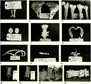
1: The 4th and 5th metacarpals belonging to the previous skeleton (T.36; S.23-43). The proximal ends (bases) of the two bones are connected together by a bar of bone, most probably ossified haematoma after trauma.
2: Outer surfaces of the right and left innominate bones of the previous skeleton (T.36; S.23-43). Notice the marked roughness and the multiple small bony osteophytes projecting from all margins of the bones.
3: Posterior surface of the upper part of femur, posterior surfaces of right and left tibiae and anterior surface of humerus belonging to the previous skeleton (T.36; S.23-43). Notice the marked roughness of the ridges and sites of muscular and ligamentous attachments.
4: Anterior surfaces of the right and left patellae of the previous skeleton (T.36; S.23-43) showing rough, irregular anterior surfaces of both bones.
5: Anterior surface of the sternum belonging to the previous skeleton (T.36; S.23-43). Notice the signs of aging as fusion of the manubrium and body, ossified xiphoid process as well as ossified costal cartilages and their fusion with the body.
6: Upper surfaces of both right and left 1st ribs belonging to the previous skeleton (T.36; S.23-43). Notice the costal cartilages that are partly ossified but not fused with the rib itself.
7: Outer surfaces of three right ribs from the previous skeleton (T.36; S.23-43) showing sites of healed fracture shafts (arrows). Notice the bony projection at the anterior end of rib (a) and the roughness of the head and neck of rib (b) which may be due to trauma and osteoarthritis.
8: A lumbar rib belonging to the previous skeleton (T.36; S.23-43). Note that the rib is short with a rounded tip.
9: Upper surface of the 7th C.V. of the previous skeleton (T.36; S.23-43). Notice the presence of a cervical rib on the right side fixed to the transverse process and the incomplete costal bar on the left side. Notice also the presence of osteophytes projecting from the anterior margin of the body of the vertebrae.
10: Anterior aspect of upper T.V. from the 2nd to the 6th belonging to the previous skeleton (T.36; S.23-43). The bodies of these vertebrae are fused together by ossified right anterior longitudinal ligament (arrows), a case of DISH.
11: Anterior aspect of the lower 6 T.V. from 7th to 12th belonging to the previous skeleton (T.36; S.23-43). These vertebrae are fused through ossified anterior longitudinal ligament on the right side (arrows). The lower 3 vertebrae, 10th, 11th and 12th are also connected by the ossified ligament of the left side (arrowhead).
12: Higher magnification of part of the previous picture to show the ossified bands of the anterior longitudinal ligament (All).
13: Anterior aspect of lumbar vertebrae belonging to the previous skeleton (T.36; S.23-43) to show the huge bony osteophytes connecting the vertebrae together.
|