| (拡大画面:129KB) |
 |
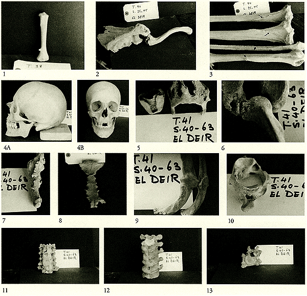
1: Tibia belonging to a child of about two years old (T.38; S.31-53). Notice the small projecting bone, this is most probably the bony part of an osteochondroma.
2: Scapula and clavicle belonging to an adult male of about 40 years old (T.40; S.25-45). Notice the region of the acromioclavicular joint which show rough irregular surfaces with cavitations and bone erosion in both the acromion and the lateral end of the clavicle; most probably it is a case of dronic osteomyelitis.
3: Bones of upper and lower limbs belonging to the previous skeleton (T.40; S.25-45). Notice the rough muscular impressions: soleal line of tibia (thin arrows), deltoid tuberosity of humerus (arrowhead) and interosseous border of the fibula (thick arrow).
4: Skull and mandible belonging to an adult male of about 45 years old (T.41; S.40-63). 1st - Lateral view to show the rounded occiput. Notice the healed factured nasal bone. 2nd - Anterior view to show the huge mandible and the deviated nasal septum.
5: Ossified thyroid and cricoid cartilages belonging to the previous specimen (T.41; S.40-63).
6: Parts of a scapula and a clavicle belonging to the previous specimen (T.41; S.40-63). The bones in the region of the acromioclavicular joint are rough, irregular with cavities, most probably a case of chronic osteomyelitis.
7: A 5th metatarsal bone belonging to the previous skeleton (T.41; S.40-63) showing marked patches of new bone formation on the surface of the bone.
8: Sternum of the previous specimen (T.41; S.40-63) showing manubrium fused with the body, ossified xiphoid process. Also there is a part of the ossified 6th costal cartilage fixed to the body of the sternum.
9: Two adjacent ribs belonging to the previous specimen (T.41; S.40-63) are seen connected together through an ossified haematoma and a callus due to trauma. A false joint is formed in the callus (pseudoarthrosis).
10: Atlas vertebra belonging to the previous specimen (T.41; S.40-63). Notice the ossified ligament which bridges across the groove on the posterior arch for the vertebral artery.
11: Cervical vertebrae from the 3rd to the 7th belonging the previous specimen (T.41; S.40-63). The vertebrae are fused by ossified anterior longitudinal ligament on both the right and the left sides (DISH).
12: Lower thoracic vertebrae belonging to the previous specimen (T.41; S.40-63). The vertebrae are joined together on the right side by large osteophytes and by ossified anterior longitudinal ligament (DISH).
13: A lumbar vertebra belong to previous specimen (T.41; S.40-63) showing a large projecting osteophytes.
|