| (拡大画面:182KB) |
 |
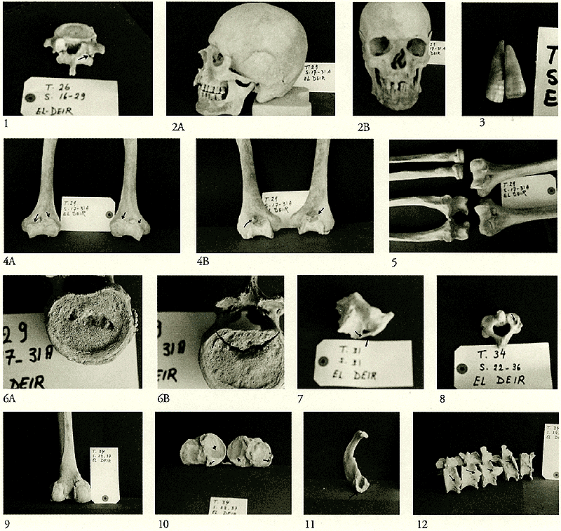
1: The 5th L.V. of the previous specimen (T.26; S.16-29) showing unilateral spondylolysis (arrow).
2: Skull belonging to an adult male of about 40 years old (T.29; S.17-31A). 1st - Lateral view: notice the flat occiput and the rounded head. 2nd - Frontal view: notice the huge mandible and the deviated nasal septum.
3: Two separate incisors from the previous specimen (T.29; S.17-31A) showing the colored striations, a condition called enamel hypoplasia.
4: Lower parts of the right and left humerus belonging to the skeleton of an adult male of about 45 years old (T.29; S.17-31A). 1st - Anterior aspect showing the lipping and the new bone formations on the articular surfaces of the trochlea and capitulum (arrows). 2nd Posterior aspect showing the new bone formations filling the olecranon fossa (arrow).
5: Parts of right and left humeri, radii and ulnae belonging to the previous skeleton (T.29; S.17-31A). Notice the roughness, lipping and bony osteophytes in the articular surfaces of the bones around the elbow region.
6: Lower T.V. belonging to the same previous skeleton (T.29; S.17-31A). 1st - The superior surface of the 10th T.V. showing a large Schmorl's node. 2nd - The superior surface of the 11th T.V. showing a large Schmorl's node in addition to a compression fissure fracture with smooth margins but no other signs of healing.
7: Part of a right scapula belonging to a young adult male of about 40 - 50 years old. (T.31; S.31) showing ossified suprascapular ligament (arrow).
8: Axis vertebra belonging to a female child of about seven years old (T.34; S.22-36). Note the presence of a rounded depression, may be a bone cyst, on the right superior articular facet (arrow).
9: Posterior aspect of the lower part of left femur belonging to a skeleton of an old male above 60 years of age (T.34; S.22-37). Notice the lipping of the margins of the articular surfaces of the femoral condyles.
10: Upper aspect of the tibial condyles belonging to the previous specimen (T.34; S.22-37) showing rough, irregular margins of the articular surfaces with bony osteophytes (arrows). Notice the marked eburnation on the medial condyles on both sides (arrowheads).
11: The right 1st rib of the previous skeleton (T.34; S.22-37) showing ossified costal cartilage (arrow).
12: Lumbar vertebrae belonging to the previous skeleton (T.34; S.22-37). Notice the large projecting osteophytes at the adjacent margins of their bodies of the vertebrae (arrows).
|