| (拡大画面:195KB) |
 |
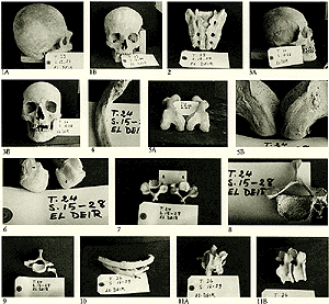
1: Skull belonging to a young adult female of about 20 years (T.22; S.12-24). 1st - Laterad view showing the round flat occiput; 2nd Anterior view.
2: Sacrum belonging to the previous specimen of the young adult female of about 20 years (T.22; S,12-24) showing spina bifida affecting almost all the sacral canal.
3: Skull and mandible belonging to an old male above 60 years of age (T.24; S.15-28), 1st - lateral view showing the flat rounded occiput. There is an obliquity of the occiput from side to side as the 2 parietal eminences are not on the same plane. 2nd - Anterior view showing the broad strong mandible.
4: Clavicle belonging to the previous case of the aged male (T.24; S.15-28). Notice the under surface of the medial part of the clavicle which is rough with a small articular facet (arrow) which most probably articulates with ossified 1st costal cartilage.
5: Right and left femora belonging to the previous specimen (T.24; S.15-28). 1st - Lower aspect of the femoral condyles of both right and left bones showing irregular patches of new bone formations. 2nd - Higher magnification showing the patches of new bone formation on the articular surfaces (arrowheads).
6: Heads of both right and left 1st metatarsal bones belonging to the previous skeleton (T.24; S.15-28) showing signs of advanced osteoarthritis in the form of eburnation (arrow heads) as well as marked lipping of the articular surfaces (arrows).
7: 2nd and 3rd C.V. belonging to the previous skeleton (T.24; S.15-28). Signs of osteoarthritis are seen in the left inferior articular facet of the 2nd C.V. (A) and in the left superior articular facet of the 3rd C.V. (B).
8: Upper aspect of the 6th C.V, of the previous specimen (T.24; S.15-28). Notice that the gap between the body and the articular mass on the right side (arrow) is narrower than that on the left side. The costal element and the foramen transversarium are absent on the right side.
9: The 7th C.V. of the previous specimen (T.24; S.15-28) showing that a separate cervical rib replaces the costal element on the left side.
10: Two ribs belonging to an adult male around 50 years of age (T.26; S.16-29). Notice the presence of a callus at the site of healed fracture shafts of both ribs (arrows).
11: Vertebrae belonging to the previous specimen (T.26; S.16-29). 1st - The 7th C.V. is seen fused with the 1st T.V. by osteophytes projecting anteriorly. 2nd - Two lower T.V. showing lipping and osteophytes.
|