|
臨床研究
Influences of Age and Gender on Results of Noninvasive Brachial-ankle
Pulse Wave Velocity Measurement-A Survey of 12517 Subjects
Hirofumi Tomiyama a, Akira Yamashina a,*. Tomio Arai a, Kenichi Hirose a,
Yutaka Koji a, Taishiro Chikamori a, Saburoh Hori b, Yoshio Yamamoto c,
Nobutaka Doba d, Shigeaki Hinohara d
Abstract
The present study was conducted to evaluate the influences of age and gender on the results of noninvasive brachial-ankle pulse wave velocity (baPWV). In 12517 subjects who had no medication and no history of cardiovascular diseases, multiple regression analysis demonstrated that age, blood pressure, body mass index, triglycerides, blood glucose, and uric acid were significant variables for baPWV in both genders. From this population, we extracted 7881‘healthy subjects’(4488 males and 3393 females, 25-87 years) without any of the atherogenic risk factors, and the results of baPWV were analyzed chronologically in 5-year age intervals. baPWV was lower in females than in males until age 60, and became similar in both genders over age 60. Multiple regression analysis demonstrated that not only the value of R2 but also the coefficient of the effect of age on baPWV are larger in females than in males. In the estimation of the regression curve, the relationship between age and baPWV demonstrated a quadratic curve in both genders. Thus, aging influences baPWV, and its effect is more prominent in female. Menopause seems to be the crucial phenomenon to explain the augmented increase in arterial stiffness with aging in females.
(c) 2002 Elsevier Science Ireland Ltd. All rights reserved.
Keywords: Pulse wave velocity; Age; Gender; Pulse pressure
1. Introduction
Increasing arterial stiffness is one of pathological states of vascular damages, and is closely associated with atherosclerotic cardiovascular diseases [1]. Pulse wave velocity (PWV) is known to be an indicator of arterial stiffness [2,3] and a marker of vascular damages [4,5]. While carotid-femoral PWV is an established method for measuring PWV [4,5], a simpler method of measuring brachial-ankle PWV (baPWV) is recently available [6,7]. We have demonstrated the validity and reproducibility of baPWV and its potential use for screening vascular damage in a large population [7].
Contrary to carotid-femoral PWV, baPWV includes peripheral components of the arterial tree. Because the influence of age differs in different parts of the arterial tree [8], a proper evaluation of the influence of age and gender on baPWV is mandatory. This information is indispensable to establish the application of baPWV as a surrogate marker in the diagnosis and management of atherosclerotic cardiovascular diseases.
The present study was conducted to evaluate the influences of age and gender on baPWV. However, previous studies have demonstrated that conventional atherosclerotic risk factors such as hypertension [9], diabetes mellitus [10], dyslipidemia [11], obesity [12], and smoking [13] influence PWV. In this study, we first verified the significance of these atherosclerotic risk factors on baPWV in over 12000 subjects. Then, to eliminate these confounding factors, we extracted a population of‘healthy subjects’who had no atherosclerotic risk factors, and analyzed the baPWV by gender and by 5-year age intervals (from age 25 to over 70).
2. Methods
2.1. Instruments
Brachial-ankle PWV was measured using a volume-plethymographic apparatus (form PWV/ABI, Colin Co. Ltd, Komaki, Japan). This instrument records PWV, blood pressure, electrocardiogram, and heart sounds simultaneously [6,7]. The subject was examined in supine position. Electrodes of electrocardiogram were placed on both wrists, a microphone for detecting heart sounds was placed on the left edge of sternum, and cuffs were wrapped on both brachia and ankles. The cuffs were connected to a plethymographic sensor that determines volume pulse form and an oscillometric pressure sensor that measures blood pressure. The pulse volume waveforms were recorded using a semiconductor pressure sensor (the sample acquisition frequency for PWV was set at 1200 Hz).
Volume waveforms for the brachium and ankle were stored for a sampling time of 10 s with automatic gain analysis and quality adjustment. Sufficient waveform data were obtained in this stored sample. McDonald reported that the mean value of the phase velocity above 2-2.5 Hz was very close to the wave front velocity [14]. The characteristic points of waveforms were determined automatically according to this phase velocity theory. The components over 5 Hz were stored using pass-filter and the wave front was determined. The time interval between the wave front of brachial waveform and that of ankle waveform was defined as the time interval between brachium and ankle (ΔTba). The distance between sampling points of baPWV was calculated automatically according to the height of the subject. The path length from the heart to the brachium (Lb) was obtained from superficial measurements and was expressed using the following equation: Lb = 0.2195 × height of the patient (cm) - 2.0734. The path length from the heart to ankle (La) was expressed using the following equation: La = (0.8129 × height of the patient (cm) + 12.328). Finally, baPWV was calculated from the following equation: baPWV = (La - Lb)/ΔTba.
The time interval between the foot of second heart sound and the dicrotic notch of brachial waveform was defined as the time interval between heart and brachium (ΔThb). Heart-brachial PWV was obtained using the following equation: heart-brachial PWV = Lb/ΔThb.
As previously mentioned [7], the coefficients of variation (CV) in reproducibility of baPWV were as follows. In healthy subjects, interobserver CV (n = 15) was 2.4% and intraobserver CV (n = 17) was 5.8%. In patients with coronary heart diseases, interobserver CV (n = 18) was 8.4% and intraobserver CV (n = 41) was 13.3%. CV in reproducibility of heart-brachial PWV measured in the same subjects as for baPWV were as follows. In healthy subjects, the interobserver (n = 15) and intraobserver (n = 17) CV were 3.1 and 9.1%, respectively. In patients with coronary heart diseases, the interobserver (n = 18) and intraobserver CV (n = 41) were 10.7 and 11.7%, respectively.
In all the studies, baPWV and heart-brachial PWV were obtained after at least 5 min rest.
2.2. Subjects
First, a total of 12517 subjects (age range: 25-87 years) were recruited from those who underwent annual health screening examinations (including the measurement of baPWV) at Tokyo Medical University Hospital or its affiliated institutes. Informed consent was obtained from all subjects. The medical history and symptoms of each subject were confirmed by the consulting doctor. All the subjects were not on medication; had no medical history of atherogenic diseases, cardiovascular diseases, renal insufficiency (serum creatinine ≧1.5 mg/dl), or other diseases requiring medical treatment; and had a normal ankle/brachial pressure index determined form PWV/ABI (>0.9). Among these subjects, 7881‘healthy subjects’with no atherosclerotic risk factors were extracted for further analysis of baPWV by gender and by age. A‘healthy subject’was defined by the following criteria: blood pressure <140/90 mmHg, fasting blood glucose <110 mg/dl, total cholesterol <240 mg/dl, triglycerides < 150 mg/dl, uric acid <7.5 mg/dl, body mass index <25, and no history of smoking.
2.3. Laboratory measurements
Plasma total cholesterol, high-density lipoprotein cholesterol, triglycerides, uric acid, and blood sugar levels were measured enzymatically. All blood samples were obtained in a fasting state in the morning.
2.4. Statistics
Data are expressed as mean ±SD. Statistical analysis was performed using the SPSS software package (SPSS, Chicago, IL). Difference between two groups were evaluated by Wilcoxon's test. Linear regression analysis was performed to evaluate the association between baPVW and other clinical variables. Then, step-wise multiple regression analysis was performed to determine the correlation and independent variables for baPWV. A value of P<0.05 was considered statistically significant.
|
Table 1 Athoropometrics in all participants (n = 12517)
|
| Gender |
Male |
Female |
P-value |
Number
Age
SBP(mmHg)
DBP(mmHg)
MBP(mmHg)
PP(mmHg)
baPWV (cm/s)
BMI (kg/m2)
TC (mg/dl)
HDL (mg/dl)
TG (mg/dl)
T/HR
FBS (mg/dl)
UA (mg/dl)
Smoke |
8227
45±11
126±15
80±10
96±12
46±8
1290±249
24±4
195±30
57±13
113+77
3.6±1.0
95±17
6.0±1.2
3097 |
4290
45±12
117±16
72±10
89±13
45±9
1166±244
21±3
200±34
73±15
71±38
2.9±0.7
91±10
4.4±0.9
515 |
ns
P<0.01
P<0.01
P<0.01
P<0.01
P<0.01
P<0.01
P<0.01
P<0.01
P<0.01
P<0.01
P<0.01
P<0.01
P<0.01 |
|
Abbreviations: SBP, systolic blood pressure; DBP, diastolic blood pressure; MBP, mean blood pressure; PP, pulse pressure; baPWV, brachial-ankle pulse wave velocity; BMI, body mass index; TC, plasma level of total cholesterol; HDL, plasma level of high-density cholesterol; T/HR, total cholesterol and high-density cholesterol ratio; TG, plasma levels of triglycerides; FBS, fasting blood glucose; UA. plasma level of uric acid; Smoke, number of subjects having history of smoking. |
3. Results
Table 1 depicts the anthropometrics of all 12517 participants.
Table 2 depicts the coefficients of correaltion in linear regression analysis between baPWV and other clinical variables in all participants. This analysis showed that hemodynamic variables and parameters reflecting either atherosclerotic risk factors or metabolic disorders correlated with baPWV in both genders. Table 3 depicts the results of step-wise multiple regression analysis between baPWV and other clinical variables in both genders. Age and blood pressure variables were potent significant variables for baPWV, and body mass index, triglycerides, uric acid, and fasting blood sugar were significant but weak variables for baPWV in both genders.
|
Table 2
|
Coefficients of correlation from linear regression analysis
between baPWV and other clinical variables in all subjects (n = 12517)
|
| Correlates |
Male |
Female |
Age
SBP
DBP
MBP
PP
BMI
TC
HDL
T/HR
TG
FBS
UA |
0.50a
0.61a
0.61a
0.64a
0.34a
0.04a
0.15a
0.03a
0.07a
0.12a
0.18a
0.12a |
0.68a
0.75a
0.68a
0.75a
0.60a
0.26a
0.34a
-0.05a
0.30a
0.33a
0.33a
0.27a |
|
Abbreviations: SBP, systolic blood pressure; DBP, diastolic blood pressure; MBP, mean blood pressure; PP, pulse pressure; BMI, body mass index; TC, plasma level of total cholesterol; HDL, plasma level of high-density cholesterol; T/HR, total cholesterol and high-density cholesterol ratio; TG, plasma levels of triglycerides; FBS, fasting blood glucose; UA, plasma level of uric acid.
a P<0.01 |
|
Table 3
|
Results of step-wise multiple regression analysis to assess
the correlation of baPWV with other variables in all subjects (n = 12517)
|
| Covariate |
Beta |
t-value |
p-value |
R2 |
| In male |
|
|
|
0.56 |
Age
SBP
DBP
MBP
PP
BMI
TC
HDL
T/HR
TG
FBS
UA
Smoke |
0.32
0.35
0.22
-0.08
0.04
0.43
0.36
|
34.13
16.45
9.82
-9.85
5.20
5.20
4.22
|
0.01
0.01
0.01Ns 0.01Ns Ns Ns 0.01
0.01
0.01Ns |
|
| In female |
|
|
|
0.70 |
Age
SBP
DBP
MBP
PP
BMI
TC
HDL
T/HR
TG
FBS
UA
Smoke |
0.37
0.39
0.19
-0.09
-0.02
0.03
0.04
0.04
|
34.79
15.08
7.17
-9.09
-2.44
3.12
4.02
4.45
|
0.01
0.01Ns 0.01Ns 0.01Ns 0.01Ns 0.01
0.01
0.01Ns |
|
|
Abbreviations: SBP, systolic blood pressure; DBP, diastolic blood pressure; MBP, mean blood pressure; PP, pulse pressure; BMI, body mass index; TC, plasma level of total cholesterol; HDL, plasma level of high-density cholesterol; T/HR. total cholesterol and high-density cholesterol ratio; TG, plasma levels of triglycerides; FBS, fasting blood glucose; UA, plasma level of uric acid; Smoke, subjects having history of smoking. |
To study the effect of gender and age on baPWV, confounding factors were eliminated by extracting a group of healthy subjects (n = 7881) free of the atherosclerotic factors. Table 4 depicts the anthropometrics of 7881 healthy subjects (4488 males and 3393 females; age: 43±11 years, range: 25-87 years) by chronological classification. While systolic and diastolic blood pressures were lower in females than in males aged <60 years, systolic blood pressure was similar in both genders from age 60. Females were shorter than males in all age groups.
|
Table 4
|
Anthropometrics in healthy subjects (n = 7881) in
chonological classification
|
| Age (yr) |
25-29 |
30-34 |
35-39 |
40-44 |
45-49 |
50-54 |
55-59 |
60-64 |
65-69 |
70- |
| Numb |
|
m
f |
158
168 |
1037
781 |
875
722 |
574
531 |
504
383 |
590
368 |
415
239 |
194
101 |
79
50 |
62
50 |
| Height (cm) |
|
m
f |
172±6
160±5a |
173±6
160±5a |
172±6
160±5a |
172±5
159±5a |
170±6
158±5a |
170±6
157±5a |
168±6
156±5a |
168±5
155±5a |
166±5
154±5a |
164±6
152±6a |
| BMI (kg/m2) |
|
m
f |
22±2
19±2a |
22±2
20±2a |
22±2
20±2a |
23±2
20±2a |
23±2
21±2a |
23±2
21±2a |
23±2
21±2a |
23±2
21±2a |
23±2
21±2a |
22±2
20±2b |
| SBP (mmHg) |
|
m
f |
119±9
108±9a |
119±9
108±9a |
119±9
109±9a |
119±9
112±9a |
120±9
114±11a |
121±9
116±10a |
122±10
119±11b |
122±9
120±11 |
123±9
122±11 |
124±11
124±11 |
| DBP (mmHg) |
|
m
f |
71±7
65±6a |
73±6
66±6a |
74±6
68±7a |
75±7
70±7a |
77±7
71±8a |
78±6
72±8a |
78±6
74±7a |
79±6
74±8a |
79±6
75±8a |
77±7
74±6a |
| HR (bpm) |
|
m
f |
63±10
64±10 |
62±9
64±9a |
64±10
67±10a |
64±9
66±9a |
64±9
66±10a |
64±9
65±9b |
63±9
66±10a |
63±9
65±8b |
64±11
68±10b |
64±11
71±10a |
| TC (mg/dl) |
|
m
f |
176±24
178±26 |
180±23
177±23b |
183±23
186±24b |
190±23
187±24b |
191±24
196±25a |
193±23
204±23a |
195±22
207±22a |
194±22
209±19a |
197±23
201±23 |
196±22
209±20a |
| HDL (mg-dl) |
|
m
f |
59±10
72±13a |
58±10
72±13a |
58±12
74±14a |
59±13
73±15a |
61±13
73±15a |
59±12
75±15a |
60±13
74±15a |
62±14
74±15a |
62±14
70±13a |
63±16
71±15a |
| T/HR |
|
m
f |
3.1±0.6
2.5±0.6a |
3.2±0.7
2.5±0.5a |
3.2±0.7
2.6±0.5a |
3.4±0.8
2.6±0.6a |
3.3±0.8
2.8±0.6a |
3.4±0.7
2.8±0.6a |
3.4±0.7
2.9±0.6a |
3.3±0.7
2.9±0.6a |
3.4±0.7
2.9±0.5a |
3.2±0.7
3.0±0.7 |
| TG (mg/dl) |
|
m
f |
77±28
54±18a |
78±29
54±23a |
83±29
57±21a |
85±29
61±21a |
85±29
68±27a |
87±30
68±25a |
87±27
73±29a |
84±24
73±25a |
81±31
76±25 |
81±28
86±28 |
| FBS (mg/dl) |
|
m
f |
89±6
86±6a |
89±7
86±6a |
92±8
89±6a |
92±8
90±7a |
93±8
91±8a |
95±9
92±7a |
95±9
93±9a |
98±10
96±10 |
97±9
94±7b |
96±8
95±9 |
| UA (mg/dl) |
|
m
f |
5.7±0.9
4.1±0.8a |
5.7±09
4.1±0.8a |
5.7±0.9a
4.1±0.8a |
5.8±0.9
4.2±0.8a |
5.8±1.0
4.2±0.8a |
5.7±1.0
4.4±0.9a |
5.6±1.0
4.5±0.9a |
5.7±1.0
4.5±0.8a |
5.9±0.9
4.6±0.8a |
5.6±0.9
5.0±1.0b |
|
Abbreviations: Numb, number of subjects; BMI, body mass index; SBP, systolic blood pressure; DBP, diastolic blood pressure; HR, heart rates; TC, plasma level of total cholesterol; HDL, plasma level of high-density cholesterol; T/HR, total cholesterol and high-density cholesterol ratio; TG, plasma levels of triglycerides; FBS, fasting blood glucose; UA, plasma level of uric acid; m, male; f, female.
a P <0.01 vs. male.
b P <0.05 vs. male. |
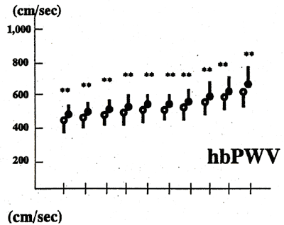
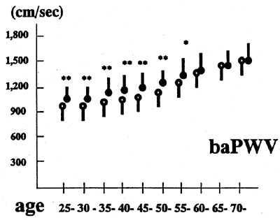
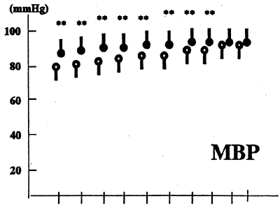
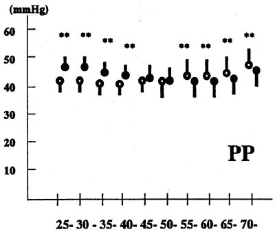
Fig. 1 depicts the chronological changes in pulse pressure, mean blood pressure, heart-brachial PWV, and baPWV. While pulse pressure was lower in females than in males until age 45, it was higher in females than in males from age 55. On the other hand, baPWV was lower in female than in male until age 60, and was similar in both genders from age 60. Heart-brachial PWV was higher in male than in female in all ages.
Table 5 depicts the results of step-wise multiple regression analysis between baPWV and other clinical variables in both genders. Age is an independent variable for baPWV. Blood pressure, but not pulse pressure, was a significant variable for baPWV in both genders. In addition, not only the value of R2 in the regression analysis, but also the coefficient of the effect of age on baPWV is larger in female than that in male. In estimation of the regression curve, the relationship between age and baPWV demonstrates a quadratic curve in both genders as follows:
Male: baPWV =0.20 × age2 - 12.13 × age + 1341.34 (R2 = 0.16, P < 0.01)
Female: baPWV = 0.16 × age2 - 4.40 × age + 977.52 (R2 = 0.37, P < 0.01)
Fig. 1.
| Chronological changes in baPWV, heart-brachial PWV, mean blood
pressure, and pulse pressure in both genders. Abbreviation: Open circle
represents female and closed circle represents male. hbPWV, heart-brachial pulse
wave velocity, baPWV, brachial-ankle pulse wave velocity, MBP, mean blood pressure,
PP, pulse pressure. *P <0.05 between
male and female, **P <0.01 between
male and female. |
a The Second Department of Internal Medicine, Tokyo Medical University.
b The Preventive Medical Center, St. Luke's International Hospital, Tokyo, Japan
c The Health Care Center, Kajima Corporation, Tokyo, Japan
d The Life Planning Center, Tokyo, Japan
『Atherosclerosis』(166 (2003) 303-309) に掲載
|