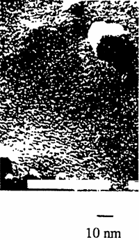
Fig. 2. A TEM photograph of minerals.
The photo shows several of these 20 A particles grouped together. These results show that the extracted minerals consisted of primary particles of about 20 A in diameter that merged to form 200 A secondary particles. As the concentration of these secondary particles increased, they in turn merged to form 1000 A particles. These larger particles then grouped together in twos And threes. Thus, these minerals exhibited a stratified structure.
The fundamental structure of each component mineral of the above rocks was tetrahedral silicate. The arrangement of elements around the apexes of the tetrahedra determined which mineral was formed. Therefore, the primary (20 A) particles of the minerals were composed of tetrahedral silicate. Furthermore, the sulfuric acid used to extract the minerals easily eluted elements such as Al, Fe, and Mg from the rock. These elements then rearranged into sulfuric ions (tetrahedra) to form alum microcrystals. In other words, the primary (20 A) particles could have been compounds of alum microcrystals and minerals composed of tetrahedral silicates.
Note that minerals are structural bodies and are not aggregates of elements. Compared with simple elements, such as Fe, Mg, Mn, the elements coordinated in the tetrahedral silicates had a catalytic power that was 104 to 105 times higher, which we easily estimated from the catalytic power of the microparticles. This amplification is called the mineral effect, which is responsible for phenomena such as photo-catalytic reaction in the growth speed of microbes.
2.3. The structure of pure water
Water structure has long been studied using neutron dispersion and X-ray radius vector distribution curves [2,3], as shown in Figs. 3 and 4, respectively.