A 1x2cm smear (200mm2) will have approximately 10000 microscopic fields if scan with light microscope using oil immersion lens (0.02mm2/microscopic field). As it is impossible to scan whole smear, the result will be issued after scanning about 300 fields. In order to issue positive results with more than 90% probability, the specimen should contain more than 30000 bacilli per ml.
Fluorochrome stained smear microscopy (FM) is suitable to a busy laboratory such as intermediate city/provincial laboratory or central laboratory where relatively large flow of specimens are processed daily. FM allows to scan at least 17 times wider microscopic field than the light microscopic field. However, FM does not permits to pick up more positives but curtails scanning time. This explains why it is irrational to use FM in a small laboratory, that requires expensive equipment and maintenance.
False positives are more often encountered among FM positives than among ZN positives. Thus it seems to be safe to report FM positives after reconfirmation by ZN method if less than 40 AFB seen.
Smear-positive culture negative results are occasionally encountered. The main reason for high rate of culture negatives among new cases is presumed to be a delayed culture in most cases. The possible explanations for false smear-positives or smear-positive culture negatives are as follows.
(1) Technical or clerical errors in smear microscopy and recording.
(2) Artefacts in the specimen, working solutions or rinse water.
(3) Cross contamination in staining.
(4) Contamination of psychropilic mycobacteria.
(5) Delayed culture of specimen left for a long period of time at high temperature.
(6) Expectoration of dead bacilli in patients treated with regimens containing rifampicin and/or with advanced cavitary disease.
(7) Harsh chemical treatment for decontamination.
The peripheral HC laboratory workers sometimes pass their works to the intermediate or central laboratory because they overestimate sensitivity of FM and of culture examination. In a study it was found that two or three smears for each patient will bring the sensitivity of sputum smear microscopy close to that of a single culture. Hence it must be emphasized to the HC workers that every efforts should be exerted to a good smear examination.
Detailed staining techniques including formula of working solutions and timing for staining, decolorization and counter-staining must be clearly described in a working manual.
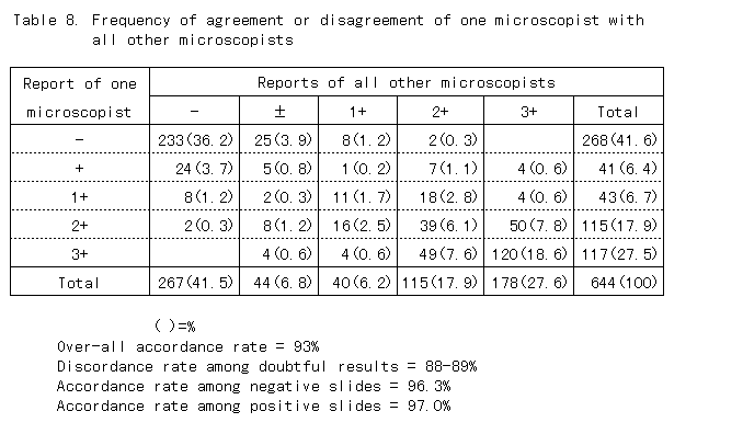
3. Reliability of smear microscopy
Unlike radiography, smear microscopy is reliable technique as seen in table 8. Agreement of microscopy results of one microscopist with others was 97% in case of positive slides and 96.3% in negative slides. Thus over-all accordance rate was 93%. However, discordance rate among doubtful results. 1-2 bacilli on a whole smear ( ) was very high, 88-89%, thus repeated examination is recommended for those cases.