|
Survey Report on the Control of Schistosomiasis in Northeast Cambodia
SUMMARY OF ACTIVITY
A survey of schistosomiasis mekongi was conducted along the Mekong river in Cambodia during the period from April 25 to May 9, 2002. The present survey focused on (1) 'rapid ELISA'-based seroepidemiological studies of schistosomiasis mekongi, (2) ultrasonographic assessment of the abdominal pathology in patients suffering from the disease, (3) collection of intermediate host snails with a view to maintaining the life cycle of the disease-causing parasite, Schistosoma mekongi, in a laboratory setting and supplying the parasite materials necessary for further surveys.
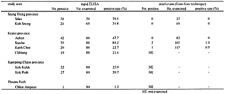
Seroepidemiological studies were conducted in eight villages along the Mekong river, targeting local schoolchildren in each study area. In the present survey, we employed an improved ELISA procedure specially designed for use in field surveys. Seropositivity rates determined in each of the study areas were 79.5% and 34.8% at Sdau and Koh Sneng (in Stung Treng province), 47.7%, 85.2%, 22.7% and 21.6% at Achen, Sanibo, Kanh Chor and Chhlong (in Kratie province) and 25.0% and 30.7% at Kok Kokh and Kok Prah (in Kampong Cham province), respectively.
Ultrasonographic examination was done to assess abdominal pathology due to S. mekongi infection in a total of 421 patients of various ages in the villages of Sdau, Koh Sneng (in Stung Treng province) and Achen, Sambor, Kahh-Chor, Chhlong, Kampong Krabei (in Kampong Cham province). Ultrasonographic images of schistosomiasis mekongi patients were unique; somewhat similar to those seen in schistosomiasis mansoni as characterized by "starry sky", "rings pipe-stems", "ruff around portal bifurcation", "patches" and "bird claw" patterns. On the other hand, no patients exhibited the "network" pattern, which is typical in patients with severe schistosomiasis japonica.
As many as 2000 intermediate host snails, Neotricula aperta, were collected at Kanpe and Krakor in Kratie province. All of the collected snails were brought back to our laboratory at the Department of Tropical Medicine and Parasitology, Dokkyo University School of Medicine. They have been yielding successful data on the life cycle of S. mekongi in a laboratory setting and providing parasite materials indispensable for further seroepidemiological surveys of the disease.
BACKGROUND
Schistosomiasis mekongi, due to a human blood-living parasite S. mekongi, has been a serious health problem in the lower Mekong Basin including Cambodia. Until recently, only little has been known about the epidemiology of the disease in Cambodia, although more than thirty years have passed since the discovery of the disease in 1968 at Kratie town, in the northeast part of the country. We have been conducting a series of surveys on schistosomiasis mekongi in Cambodia since 1997 in collaboration with the National Malaria Center (CNM), Ministry of Health, Cambodia, for the purpose of clarifying the current epidemiological status of Mekong schistosomiasis. These surveys have revealed that highly endemic areas exist mainly in the upstream area of the Mekong river and in Kratie province, whereas the disease endemicity gradually decreases further downriver.
OBJECTIVES
The main objectives of the present survey are as follows:
1) to perform a seroepidemiological study based on a newly developed rapid ELISA technique, with which we can perform serological tests for schistosomiasis mekongi infection using only a small amount of whole blood from each subject within a short time.
2) to accumulate data on abdominal ultrasonographic images of schistosomiasis mekongi patients with a view to establishing a standard for the ultrasonographic diagnosis of the disease.
3) to collect N. aperta, the intermediate host snail of S. mekongi, for the purpose of establishing the life cycle of the parasite in a laboratory setting and obtaining the parasite materials needed for future surveys.
TEAM MEMBERS
The survey team comprised Dr. Suon Siela and two well trained medical technicians from CNM, Phnom Penh, Professor Hajime Matsuda and Dr. Jun Matsumoto from Dokkyo University School of Medicine, and Dr. Hiroshi Ohmae from Tsukuba University School of Medicine, Japan. These members received technical support from local staff belonging to the Provincial Health Department in each study area (Stung Treng, Kratie and Kampong Cham provinces).
SCHEDULE
The period of our survey was from April 25 to May 9, 2002. The schedule of the present survey is briefly shown below.
| April 25: |
Briefing at CNM, Phnom Penh |
| April 26: |
Briefing at Stung Treng Provincial Health Department |
| April 27-28: |
Sero-epidemiologic and ultrasonographic studies in Stung Treng province |
| April 30: |
Briefing at Kratie Provincial Health Department |
| May 1-5: |
Sero-epidemiological and ultrasonographic studies, and snail collection |
| May 6: |
Briefing at Kampong Cham Provincial Health Department |
| May 7-8: |
Sero-epidemiological survey in Kampong Cham province |
| May 9: |
Rebriefing at CNM, Phnom Penh |
ACTIVITIES AND RESULTS
1. Sero-epidemiological survey
In the present survey, a newly developed simple method, named 'rapid ELISA', was used in serological diagnosis of schistosomiasis mekongi infection. This serodiagnostic technique requires less than 20μL of whole blood collected from the subject's finger and does not need any electrical equipment. In this survey, the simple and rapid procedure for this ELISA technique enabled us to determine the seropositivity within an hour or so before passing on the serodiagnosis results to examinees and local working staff. One more notable improvement in the ELISA procedure is the use of S. mekongi egg antigen. Until the previous survey last year, we had alternatively used S. japonicum egg antigen because S. mekongi antigen was not available. With intermediate host snails collected in the previous survey last year, we established the S. mekongi life cycle in our laboratory and obtained the parasite material for the preparation of the egg antigen. Seroepidemiological surveys were done in eight villages along the Mekong river. A total of 636 blood samples were donated from schoolchildren, as shown in the Table. Blood specimens were collected easily by finger pricking just before performing a rapid ELISA for detecting the S. mekongi-specific antibody. Samples showing an optical density (OD) of more than 0.35 were considered positive.
The results of the serological tests, summarized in the Table, were shown in situ to study subjects and local working staff. Seropositivity rates were 79.5% (31/39) and 34.8% (24/69) at Sdau and Koh Sneng (in Stung Treng province), 47.7% (42/88), 85.2% (75/88), 22.7% (20/88) and 21.6% (19/88) at Achen, Sambo, Kanh Chor and Chhlong (in Kratie province) and 25.0% (22/88) and 30.7% (27/88) at Kok Kokh and Kok Prah (in Kampong Cham province), respectively.
The rather high seropositivities determined for children in Kok Kokh and Kok Prah are noteworthy because no case of schistosomiasis mekongi has so far been reported in Kampong Cham province, where we found no snail focus in the Mekong river. However, we should be cautious in interpreting these results in Kampong Cham province; it cannot yet be concluded that there is a hidden endemic focus of schistosomiasis mekongi in this area until patients showing positive results in parasitological examinations (i.e., stool examination) are found because the possibility of false positivity still cannot be excluded in any serological examination.
Stool examinations employing the Kato-Katz technique were also applied to the same study subjects as those seen in the seroepidemiological studies (Table 1). Only three (one in Kanh Chor and two in Sambo) out of 406 schoolchildren were found to be schistosome egg-positive in spite of the seroepidemiology results showing rather high positivity in each study area. For the last few years, large-scale mass treatment has been conducted annually by the National Malaria Center (CNM), coving all the disease endemic areas in Cambodia. This operation may have been responsible for the remarkable decrease in the number of egg-positive patients.
2. Ultrasonography
A total of 421 patients of various ages were subjected to abdominal ultrasonographic examination in Sdau and Koh-Sneng (in Stung Treng province, n = 96) and in Achen, Sambor, Kanh-Chor, Chhlong and Kampong Krabei (in Kratie province, n = 325).
We found that ultrasonographic images of liver pathology in schistosomiasis mekongi infection appeared somewhat similar to those in schistosomiasis mansoni infection as characterized by "starry sky", "rings pipe-stems", "ruff around portal bifurcation", "patches" and "bird claw" patterns. On the other hand, the "network" pattern, a typical ultrasonographic image of severe schistosomiasis japonica, was not observed even in patients with severe infection. In experimental pathologic studies using S. mekongi-infected mice, we observed that the histopathology of schistosomiasis mekongi is quite different from that of schistosomiasis japonica, even though the disease-causative agents of both diseases are closely related species. In the liver and intestine of S. mekongi-infected mice, the fibrotic changes due to parasite egg deposition in the tissues progress much more slowly than those in S. japonicum-infected mice, although there was a unique histopathological appearance resembling vacuolar generation in the host tissues. These findings, together with those of clinical ultrasonography, suggest that the pathology due to S. mekongi is unique and differs from those caused by other related Schistosoma species. Therefore, standardization of the ultrasonographic diagnosis of schistosomiasis mekongi is urgently needed based upon the accumulated data of both clinical and experimental studies.
3. Collecting intermediate host snails
In the districts of Kanpe and Krakor in Kratie province, there were a number of intermediate host snails sticking to rocks in shallows of the Mekong River. We collected about 2000 of these snails and brought them back to our laboratory at Dokkyo University School of Medicine, Japan. These snails have been artificially infected with S. mekongi and kept in water tanks, which have been yielding the parasite materials used for further research and surveys on schistosomiasis mekongi. Intensive studies are currently underway to clarify the biochemical features of the parasite antigen and its reactivity against sera from schistosomiasis patients, which should provide valuable information for development of improved serodiagnostic techniques for the diagnosis of S. mekongi infection.
| Table 1. |
Results of epidemiological survey of schistosomiasis mekongi along the Mekong river in Cambodia, 2002. |
| (拡大画面:11KB) |

|
|