However, the rest of the flows collides with each other at the tip of the needle valve and a lot of small scale vortexes are produced in the sac chamber. After t = 82 ms, cavitation bubbles in the discharge hole grow into the thin films. They develop into the sac chamber and are linked to each other. The cavitation films are supposedly produced in the boundary between the high velocity flow from the needle seat to the discharge hole and the disturbed flow in the sac chamber. The flow pattern in the sac chamber after t = 62 ms becomes approximately steady flow.
The spray behaviors for the STD Model III shown in Fig.9 have a close relation to the internal flow characteristics described above. In the initial stage of the injection until t = 8 ms, the liquid column is pushed out of the discharge hole. Around t = 14 ms, a disturbed spray issues and passes over the initially issued liquid column. Then the spray has a large spray angle with fine droplets around t = 36 ms. After t = 50 ms, the spray angle becomes a little smaller than before, but is still much larger than the spray angle for the STD Model I. Such wide spray angles for STD Model III are supposedly due to the disturbed flow in the sac chamber which is not seen in the sac chamber of the STD Model I. At t = 64 ms, the dense jet appears in the region near the discharge hole due to the bounce of the needle valve. After t = 84 ms, the spray angle fluctuates when the cavitation films flow out of the discharge hole.
丂
3.3 Mini Sac Model
The flow patterns in the sac chamber, the cavitation behaviors in the sac chamber and the discharge hole, and the spray behaviors for the Mini Sac Model are shown in Figs. 10, 11 and 12, respectively. In Fig.10 at t = 4 ms from the beginning of the needle valve opening, the flow pattern in the sac chamber is similar to the flow in the sac chamber of the STD Model III. The smooth flows from the bottom of the sac chamber to the discharge hole are seen as shown by arrows in the figure. Around t = 18 ms, the very bright portions appear around the inlets of the discharge holes, which are due to the cavitation films in the sac chamber.
This is more clearly seen in Fig.11. At t = 18 ms, the cavitation films occur in the sac chamber and then disappear. After a short interval, the spiral air cavities occur at the exit of the discharge holes and develop in the upstream direction of the flow at t = 25 ms. Around t = 28 ms to 33 ms, these spiral air cavities attach to the needle valve, are linked to each other, fully develop in the sac chamber and take a twisted thread-like shape. After t = 55 ms, the spiral air cavities change into the thin cavitation films, shrink in the sac chamber and remain mainly around the inlet of the discharge holes. Then the thin cavitation films repeat developing into the sac chamber, linking to each other, and shrinking in the sac chamber. At t = 62 ms in Fig.10, the flow pattern in the sac chamber is similar to that in the sac chamber of the STD Model I. The flows become relatively smooth and the flows from the needle seat enter the discharge holes directly. The spray behaviors for the Mini Sac Model are shown in Fig.12. The spray behaviors until t = 20 ms is almost the same as STD Model I and III. However after t = 36 ms when the spiral air cavities develop in the sac chamber, the spray shows a hollow cone structure with a large spray angle and fine droplets. The hollow cone structure of the spray continues until t = 50 ms. After t = 50 ms, the spray angle becomes a little smaller, since the spiral air cavities in the sac chamber shrink and change into the thin cavitation films in the discharge hole, though the spray angle is still larger than that of the STD Model I and III. The spray behavior after t = 64 ms is similar to that of the STD Model III.
丂
丂
4. SPRAY ANGLE VARIATIONS DURING NEEDLE VALVE OPENING AND CLOSING
丂
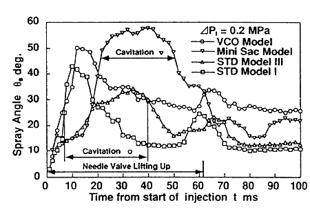
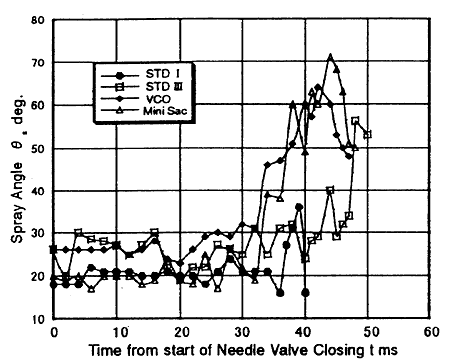
Variations of the spray angles in the opening and closing processes of the needle valve are shown in Figs. 13 and 14, respectively. The spray angle was defined as an angle between two lines connecting the outlet of the discharge hole and two edges of the spray image at a distance of 10 mm from the nozzle, 5 times of the discharge hole diameter.
In the valve opening process shown in Fig.13, the spray angles for all model nozzles increase in the initial stage of the injection, take maximums and gradually decrease. The spray angles take small maximums again when the needle valve reaches the full lift. Among the models, the Mini Sac Model takes the largest maximum spray angle, since the swirling flow occurs in the discharge hole. The largest spray angle for the VCO model is the second to that of the Mini Sac Model and the STD Model III is the third. These are supposedly due to the disturbed flow in the upstream of the discharge hole for the VCO Model and in the sac chamber flow for the STD Model III. The STD Model I shows the smallest spray angle due to the most smooth flow in the sac chamber among the models.
The spray angle variations in the needle valve closing process are shown in Fig.14. The spray angles for all models increase just before the closure of the needle valve, take maximums and suddenly decreases. Among the models, the Mini Sac Model takes the largest maximum spray angle. The largest spray angle for the VCO Model is the second to that for the Mini Sac Model, the STD Model III is the third.
丂